

Several Staphylococcus spp, while typically present in healthy poultry, can cause localized or systemic infection if skin or mucous membrane barriers are compromised. Clinical signs vary depending on the location infected. Diagnosis is confirmed by culture of lesions. Antimicrobial treatment of systemic infection is often successful; however, localized lesions can be more difficult to treat.
Staphylococcosis is a bacterial disease caused by gram-positive cocci of the genus Staphylococcus. S aureus and other species are ubiquitous in the environment and are part of normal skin microbiota and mucous membranes of poultry and other animals.
Staphylococcus spp can cause disease when they have access to tissues and the bloodstream after physical barriers (eg, skin and mucous membranes) are compromised. These bacteria also cause disease in immunosuppressed birds.
Staphylococcosis starts with a localized infection at the entry point and can become systemic. After acute bacteremia/systemic infection, bacteria can settle in different organs, resulting in additional localized lesions. Therefore, there are two major categories of staphylococcosis: systemic infection and localized lesions.
Septicemia and multiorgan infections are examples of systemic staphylococcal infections. However, the most common form of staphylococcosis is localized lesions. Examples include arthritis, tenosynovitis, osteomyelitis, and omphalitis. See the Staphylococcus spp Associated With Infections in Poultry.
Staphylococcus spp Associated With Infections in Poultry
Staphylococcus Species | Affected Poultry Species and Production Type | Systemic or Local and Associated Diseases |
|---|---|---|
S agnetis | Broiler chicken, broiler breeder, laying hen | Systemic: septicemia, endocarditis Local: bacterial chondronecrosis with osteomyelitis, focal ulcerative dermatitis syndrome |
S aureus | All poultry species and production types | Systemic: septicemia, turkey osteomyelitis complex (green liver-osteomyelitis syndrome) Local: arthritis, gangrenous dermatitis, synovitis, chondronecrosis, osteomyelitis, bumblefoot, omphalitis |
S auricularis | Broiler chicken | Systemic infection |
S capitis | Broiler chicken, laying hen, broiler breeder | Systemic infection |
S chromogenes | Broiler chicken, turkey, laying hen, broiler breeder, waterfowl | Systemic infection |
S cohnii urealyticus | Broiler chicken, turkey, laying hen, broiler breeder, waterfowl | Systemic infection Local: arthritis, scabby hip syndrome |
S epidermidis | Broiler chicken, laying hen, waterfowl | Systemic infection Local: chondronecrosis with osteomyelitis, scabby hip syndrome |
S gallinarum | Broiler chicken | Systemic infection |
S hominis | Broiler chicken | Systemic infection Local: chondronecrosis with osteomyelitis |
S hyicus | Broiler chicken, laying hen, broiler breeder, turkey | Systemic infection Local: fibrinopurulent blepharitis and conjunctivitis, stifle joint osteomyelitis, folliculitis and epidermitis, pododermatitis, dermatitis (associated with fowlpox), beak necrosis |
S intermedius | Broiler chicken | Systemic infection Local: scabby hip syndrome |
S lentus | Broiler chicken, turkey, broiler breeder, laying hen, waterfowl | Systemic infection Local: scabby hip syndrome |
S saprophyticus | Broiler chicken, turkey, laying hen | Systemic infection Local: chondronecrosis with osteomyelitis |
S sciuri | Broiler chicken, turkey, waterfowl | Systemic infection Local: scabby hip syndrome |
S simulans | Broiler chicken, turkey, waterfowl | Systemic: systemic infection, endocarditis Local: scabby hip syndrome |
S warneri | Broiler chicken | Local: scabby hip syndrome |
S xylosus | Broiler chicken, turkey, broiler breeder, waterfowl | Systemic infection Local: chondronecrosis with osteomyelitis |
Table adapted from Szafraniec GM, Szeleszczuk P, Dolka B. A review of current knowledge on Staphylococcus agnetis in poultry. Animals (Basel). 2020;10(8):1421. doi:10.3390/ani10081421 | ||
Economic losses result from decreased weight gain, death, and condemnation at slaughter.
Etiology and Pathogenesis of Staphylococcosis in Poultry
The genus Staphylococcus contains dozens of species, but S aureus is the most pathogenic. S aureus is a gram-positive, catalase-positive, coccoid bacterium that appears in grapelike clusters on stained smears (see ).
Once in the bloodstream, Staphylococcus can produce systemic disease or localized lesions in tissues. S aureus can invade metaphyses, leading to arthritis and osteomyelitis.


Staphylococcus aureus showing gram-positive staining from a pure S aureus isolate. Gram stain. Original magnification, 10,000X.
Courtesy of Dr. Mohamed M. El-Gazzar.
Most pathogenic strains have been coagulase-positive; however, coagulase-negative Staphylococcus spp (including S hyicus, S epidermidis, S simulans, and S gallinarum) have been reported from clinical cases.
Epidemiology and Transmission of Staphylococcosis in Poultry
Skin wounds, minor surgical procedures (ie, beak, toe, or comb trimming), vaccine injections, and compromised intestinal mucosa can introduce Staphylococcus into local tissue or the bloodstream. Infection can also occur in the hatchery from contamination of an open navel.
Clinical Signs and Lesions of Staphylococcosis in Poultry
Localized Infection
Bumblefoot (pododermatitis) is more common in heavier birds and in males. Swollen footpads and limping are common clinical signs.
Bumblefoot is often precipitated by injuries that allow contamination of subcutaneous tissue in the footpad. This leads to acute necrotic inflammation, which later develops into a mass of dead tissue. Microscopically, edema, necrosis, and granulomas that may contain bacterial colonies may be observed in lesions. (See photographs of , , , , , , and . See also photomicrographs of , , and .)


Swollen footpad of a chicken with bumblefoot. Note the marked subcutaneous edema evident on cut section.
Swollen footpad of a chicken with bumblefoot. Note the marked subcutaneous edema evident on cut section.
Courtesy of Dr. Mohamed M. El-Gazzar.


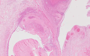
Photomicrograph of a histological section of edematous and necrotic footpad of a bird with acute bumblefoot (pododermatitis). H&E stain. Original magnification, 40X.
Photomicrograph of a histological section of edematous and necrotic footpad of a bird with acute bumblefoot (pododermat
Courtesy of Dr. Mohamed M. El-Gazzar.


Young chick with an infected yolk sac (omphalitis).
Young chick with an infected yolk sac (omphalitis).
Courtesy of Dr. Yuko Sato.


Photomicrograph of a histological section of yolk sac from a chick with omphalitis. Note the necrotic tissue, visible bacterial colonies (black arrows), and degenerating heterophils (blue arrow). These are surrounded by multinucleated giant cells (red arrow), macrophages, heterophils, and fibrous connective tissue (granuloma; green arrow). H&E stain. Original magnification, 200X.
Photomicrograph of a histological section of yolk sac from a chick with omphalitis. Note the necrotic tissue, visible b
Courtesy of Dr. Mohamed M. El-Gazzar.


Necropsy photograph of a bird with caseous tenosynovitis. Note the caseous inflammation of digital flexor tendons and tendon sheaths.
Necropsy photograph of a bird with caseous tenosynovitis. Note the caseous inflammation of digital flexor tendons and t
Courtesy of Dr. Mohamed M. El-Gazzar.
Photomicrograph of a histological section of digital flexor tendon sheath from a bird with caseous tenosynovitis. Note the edema (black arrow), congestion, hemorrhage (blue arrow), necrosis, and visible bacterial colonies (red arrow). H&E stain. Original magnification, 40X.
Photomicrograph of a histological section of digital flexor tendon sheath from a bird with caseous tenosynovitis. Note
Courtesy of Dr. Mohamed M. El-Gazzar.


Swollen shanks in a 14-week-old turkey tom, indicating inflammation of digital flexor tendons and their synovial sheaths.
Swollen shanks in a 14-week-old turkey tom, indicating inflammation of digital flexor tendons and their synovial sheath
Courtesy of Dr. Mohamed M. El-Gazzar.


Necropsy photograph of the proximal tibiotarsus of a bird with osteomyelitis. Note the extensive necrosis.
Necropsy photograph of the proximal tibiotarsus of a bird with osteomyelitis. Note the extensive necrosis.
Courtesy of Dr. Mohamed M. El-Gazzar.


Swollen joint containing excessive synovial fluid.
Swollen joint containing excessive synovial fluid.
Courtesy of Dr. Mohamed M. El-Gazzar.

Necrotic dermatitis of breast and thigh skin in a laying hen infected with Staphylococcus.
Necrotic dermatitis of breast and thigh skin in a laying hen infected with Staphylococcus.
Courtesy of Dr. Mohamed M. El-Gazzar.
Swollen footpad of a chicken with bumblefoot. Note the marked subcutaneous edema evident on cut section.
Swollen footpad of a chicken with bumblefoot. Note the marked subcutaneous edema evident on cut section.
Courtesy of Dr. Mohamed M. El-Gazzar.
Photomicrograph of a histological section of edematous and necrotic footpad of a bird with acute bumblefoot (pododermatitis). H&E stain. Original magnification, 40X.
Photomicrograph of a histological section of edematous and necrotic footpad of a bird with acute bumblefoot (pododermat
Courtesy of Dr. Mohamed M. El-Gazzar.
Young chick with an infected yolk sac (omphalitis).
Young chick with an infected yolk sac (omphalitis).
Courtesy of Dr. Yuko Sato.
Photomicrograph of a histological section of yolk sac from a chick with omphalitis. Note the necrotic tissue, visible bacterial colonies (black arrows), and degenerating heterophils (blue arrow). These are surrounded by multinucleated giant cells (red arrow), macrophages, heterophils, and fibrous connective tissue (granuloma; green arrow). H&E stain. Original magnification, 200X.
Photomicrograph of a histological section of yolk sac from a chick with omphalitis. Note the necrotic tissue, visible b
Courtesy of Dr. Mohamed M. El-Gazzar.
Necropsy photograph of a bird with caseous tenosynovitis. Note the caseous inflammation of digital flexor tendons and tendon sheaths.
Necropsy photograph of a bird with caseous tenosynovitis. Note the caseous inflammation of digital flexor tendons and t
Courtesy of Dr. Mohamed M. El-Gazzar.
Photomicrograph of a histological section of digital flexor tendon sheath from a bird with caseous tenosynovitis. Note the edema (black arrow), congestion, hemorrhage (blue arrow), necrosis, and visible bacterial colonies (red arrow). H&E stain. Original magnification, 40X.
Photomicrograph of a histological section of digital flexor tendon sheath from a bird with caseous tenosynovitis. Note
Courtesy of Dr. Mohamed M. El-Gazzar.
Swollen shanks in a 14-week-old turkey tom, indicating inflammation of digital flexor tendons and their synovial sheaths.
Swollen shanks in a 14-week-old turkey tom, indicating inflammation of digital flexor tendons and their synovial sheath
Courtesy of Dr. Mohamed M. El-Gazzar.
Necropsy photograph of the proximal tibiotarsus of a bird with osteomyelitis. Note the extensive necrosis.
Necropsy photograph of the proximal tibiotarsus of a bird with osteomyelitis. Note the extensive necrosis.
Courtesy of Dr. Mohamed M. El-Gazzar.
Swollen joint containing excessive synovial fluid.
Swollen joint containing excessive synovial fluid.
Courtesy of Dr. Mohamed M. El-Gazzar.
Necrotic dermatitis of breast and thigh skin in a laying hen infected with Staphylococcus.
Necrotic dermatitis of breast and thigh skin in a laying hen infected with Staphylococcus.
Courtesy of Dr. Mohamed M. El-Gazzar.
Omphalitis, or yolk sac infection, is a common disease caused by S aureus in young chicks and poults. Open navels can become contaminated and infected in young hatchlings and elevate mortality rates.
Chicks with omphalitis have moist, dark navels, and affected birds are often lethargic. When yolk sacs in developing chicks become infected, they are not resorbed normally within the first week of life but are retained longer. Infected yolks are abnormally colored (dark green to brown), doughy, and malodorous.
Arthritis, synovitis, and osteomyelitis are common staphylococcal infections in poultry. They can begin in joints, tendons, or bones through systemic infection or through local injury.
Clinical signs include swollen, hot joints with limping and reluctance to walk.
Affected joints contain increased synovial fluid, tendons and tendon sheaths can be inflamed, and bones can contain focal areas of necrosis, known as osteomyelitis. The femoral head and the proximal head of the tibiotarsus are commonly affected. Vertebrae can be affected as well, leading to spondylitis.
Systemic Infection
Research is limited regarding acute septicemic Staphylococcus infections. They occur in laying hens, particularly those older than 85 weeks. Clinical signs include the following:
sudden death
sharp increase in mortality rate, approaching 1% per day
sharp decrease in egg production
cutaneous inflammation and necrosis of comb, wattles, and skin overlying breast and thigh
Dead birds show lesions of acute septicemic infection, including liver necrosis; enlarged, mottled spleen; and petechial hemorrhages in brain, lungs, and proventricular glands.
Typically, the number of deaths subsides after the flock is treated with antimicrobials for 5–7 days. The morbidity rate is not high (5–10%); however, birds that are affected usually die.
See images of , , and ; see also photomicrographs of and .


Bird with septicemic Staphylococcus. Note the swollen, edematous face and wattles.
Bird with septicemic Staphylococcus. Note the swollen, edematous face and wattles.
Courtesy of Dr. Mohamed M. El-Gazzar.


Layer with septicemic Staphylococcus. Note the necrotic, hemorrhagic wattles.
Layer with septicemic Staphylococcus. Note the necrotic, hemorrhagic wattles.
Courtesy of Dr. Mohamed M. El-Gazzar.

Photomicrograph of necrotic dermatitis in the wattle of a bird with septicemic Staphylococcus. H&E stain. Original magnification, 100X.
Photomicrograph of necrotic dermatitis in the wattle of a bird with septicemic Staphylococcus. H&E stain. Original magn
Courtesy of Dr. Mohamed M. El-Gazzar.


Necrotic wattle tissue showing gram-positive cocci. Staphylococcus aureus was isolated from this tissue. Gram stain. Original magnification, 400X.
Necrotic wattle tissue showing gram-positive cocci. Staphylococcus aureus was isolated from this tissue. Gram stain. Or
Courtesy of Dr. Mohamed M. El-Gazzar.


Gross pathology photograph of the liver from a chicken with septicemia. Note the green hepatic discoloration.
Gross pathology photograph of the liver from a chicken with septicemia. Note the green hepatic discoloration.
Courtesy of Dr. Yuko Sato.
Bird with septicemic Staphylococcus. Note the swollen, edematous face and wattles.
Bird with septicemic Staphylococcus. Note the swollen, edematous face and wattles.
Courtesy of Dr. Mohamed M. El-Gazzar.
Layer with septicemic Staphylococcus. Note the necrotic, hemorrhagic wattles.
Layer with septicemic Staphylococcus. Note the necrotic, hemorrhagic wattles.
Courtesy of Dr. Mohamed M. El-Gazzar.
Photomicrograph of necrotic dermatitis in the wattle of a bird with septicemic Staphylococcus. H&E stain. Original magnification, 100X.
Photomicrograph of necrotic dermatitis in the wattle of a bird with septicemic Staphylococcus. H&E stain. Original magn
Courtesy of Dr. Mohamed M. El-Gazzar.
Necrotic wattle tissue showing gram-positive cocci. Staphylococcus aureus was isolated from this tissue. Gram stain. Original magnification, 400X.
Necrotic wattle tissue showing gram-positive cocci. Staphylococcus aureus was isolated from this tissue. Gram stain. Or
Courtesy of Dr. Mohamed M. El-Gazzar.
Gross pathology photograph of the liver from a chicken with septicemia. Note the green hepatic discoloration.
Gross pathology photograph of the liver from a chicken with septicemia. Note the green hepatic discoloration.
Courtesy of Dr. Yuko Sato.
Lesions that are sequelae of systemic infections are sometimes the only pathological findings in staphylococcal infections. Gangrenous dermatitis is observed, particularly in immunocompromised chickens, such as those affected by infectious bursal disease or chicken anemia virus infection. Gangrenous dermatitis is often due to combined infection with S aureus and Clostridium septicum and/or Escherichia coli. Affected areas are usually hemorrhagic and crepitant.
Endocarditis can also occur after systemic staphylococcal infections.
Staphylococcal septicemia can result in green liver or liver with multifocal necrotic granulomas. S aureus has been implicated as the etiological agent behind turkey osteomyelitis complex (also called green liver-osteomyelitis syndrome), which causes green discoloration of the liver and inflammatory lesions in bones, joints, or periarticular soft tissues. Green livers are common in turkeys and lead to increased condemnation in the processing plant.
Hemorrhagic enteritis virus, another immunosuppressive agent, is thought to contribute to increased frequency of green liver in turkeys.
Diagnosis of Staphylococcosis in Poultry
Clinical evaluation
Elimination of other primary pathogens
Bacterial culture
Although some lesions may be suggestive of Staphylococcus infection, diagnosis is confirmed by identifying organisms from lesions cultured on blood agar plates. Phenotyping and genetic techniques have been used to classify strains of poultry S aureus. Differential diagnoses include infection with Streptococcus spp, E coli, and Pasteurella multocida, as well as other bacterial diseases of poultry.
Staphylococcus bacteria are ubiquitous in the environment and cause disease as secondary pathogens, so mere isolation of Staphylococcus does not necessarily constitute a disease diagnosis. In fact, Staphylococcus spp are more often isolated as contaminants.
Treatment and Prevention of Staphylococcosis in Poultry
Antimicrobials for systemic infections
Improved management to prevent injuries
Staphylococcosis can be successfully treated with antimicrobials; however, a susceptibility test should be performed. Antimicrobials used to treat Staphylococcus infections include penicillin, erythromycin, lincomycin, and spectinomycin. Antimicrobials are typically administered in the sole source of drinking water according to label instructions. Appropriate egg and meat withdrawal periods must be assigned and followed.
Proper management to avoid injury and immunosuppression helps prevent staphylococcosis. Because wounds are the primary route by which Staphylococcus enters the body, decreasing potential sources of injury to birds via the following precautions is important:
Eliminating wood splinters in litter, protruding wires from cages, fighting, and cannibalism can prevent the development of skin wounds and secondary staphylococcosis.
Because beak and toe trimming in young chickens and turkeys can result in staphylococcal septicemia, ensuring that equipment is sanitary will help prevent outbreaks.
Good litter management is important in controlling footpad injuries to prevent bumblefoot.
Hatchery sanitation and good egg management practices can decrease navel infections and omphalitis.
Zoonotic Risk of Staphylococcosis in Poultry
S aureus can cause food poisoning in humans. Enterotoxin-producing strains are found on apparently healthy poultry, so proper precautions should be taken when handling and cooking poultry products.
Methicillin-resistant S aureus (MRSA) has been isolated from poultry meat in a number of countries; however, its prevalence and importance for human health are not completely understood.
Key Points
Environmental bacteria, normal flora, and opportunistic pathogens lead to staphylococcal infections in poultry.
Infections can be either systemic or localized.
Staphylococcal bacteria isolated from bacterial culture are not always the cause of clinical disease.
Antimicrobial treatment is more successful for systemic than localized infection.
For More Information
Andreason CB. Staphylococcosis. In: Swayne DE, ed. Boulianne M, Logue CM, McDougald LR, Nair V, Suarez DL, et al, associate eds. Diseases of Poultry. 14th ed. Wiley Blackwell; 2020:995-1002,
Shivaprasad HL. Staphylococcosis. In: Brugere-Picoux J, Vaillancourt JP, Shivaprasad HL, Venne D, Bouzouaia M, eds. Manual of Poultry Diseases. Association francaise pour l'avancement des sciences; 2015:374-375.
Szafraniec GM, Szeleszczuk P, Dolka B. A review of current knowledge on Staphylococcus agnetis in poultry. Animals (Basel). 2020;10(8):1421.
Meyer M, Bobeck E, Sato Y, El-Gazzar M. Comparison of naturally occurring vs. experimental infection of Staphylococcus aureus septicemia in laying hens in two different age groups. Avian Dis, 2021;65(2):310-320.


