


Balanoposthitis is inflammation of the penile or preputial mucosa. The characteristic clinical sign is excessive preputial discharge, often accompanied by signs of pain. Cytological evaluation, bacterial culture, and ultrasonography can be helpful for diagnosis. The penis and preputial cavity should be flushed and cleaned with a mild antiseptic solution; underlying predisposing factors should be treated.
Balanoposthitis, or inflammation of the penile or preputial mucosa, occurs primarily in dogs. It is rare in cats but can be associated with persistent frenula or a constricting band of hair.


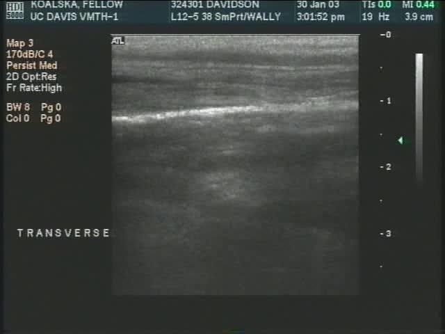
Courtesy of Dr. Autumn Davidson.
Etiology of Balanoposthitis in Dogs and Cats
Balanoposthitis can be primary, as with idiopathic lymphoplasmacytic inflammation (see primary idiopathic balanoposthitis image), or secondary, as with the following:
bacterial overgrowth
penile trauma (see fractured os penis video)
penile or preputial neoplasia (see lymphosarcoma image)
foreign bodies, such as grass awns (see hemorrhagic tissue image)


Courtesy of Dr. Autumn Davidson.


Courtesy of Dr. Autumn Davidson.


Courtesy of Dr. Autumn Davidson.
Clinical Findings of Balanoposthitis in Dogs and Cats
Dogs with balanoposthitis produce excessive, often mucopurulent, preputial discharge that can accumulate on the pelvic limbs and be associated with excessive preputial licking and signs of pain. Normal preputial secretions usually do not result in overt clinical signs. A slight mucoid preputial discharge is present in many sexually mature dogs and is of little clinical importance.
Painful swelling of the prepuce is rarely present in balanoposthitis, except in cases of trauma, snake bite, or foreign bodies.
If clinical signs of systemic illness are present, a more serious concomitant disorder should be considered.
Diagnosis of Balanoposthitis in Dogs and Cats
In patients with suspected balanoposthitis, the penis and prepuce should be thoroughly examined to the level of the fornix for underlying predisposing factors. Infusion of saline solution (0.9% NaCl) through a rigid endoscope facilitates this examination (see normal preputial fornix image), but an otoscope can be used if necessary. Sedation or general anesthesia may be needed.
Foreign bodies can be submucosal and difficult to see, so tiny tracts should be explored.
Cytological evaluation of preputial smears may be helpful. Cytological evaluation or biopsy of mucosal lesions may be indicated.
Bacterial cultures of the preputial cavity, although sometimes difficult to interpret because of the presence of normal preputial flora, may help identify dysbiosis (evidenced by growth of abnormal bacterial species or monobacterial growth on culture) and determine antimicrobial susceptibility for refractory cases.
Ultrasonography can facilitate the discovery of foreign body tracts, penile trauma (eg, hematoma, fractured os penis), or masses.
Treatment of Balanoposthitis in Dogs and Cats
Treatment of balanoposthitis includes correcting any predisposing factors, clipping long hair away from the preputial orifice to facilitate hygiene, and thoroughly flushing the preputial cavity with a mild, dilute antiseptic (eg, dilute povidone-iodine or chlorhexidine) or sterile saline solution (0.9% NaCl).
If bacterial infection is suspected, an antimicrobial ointment can be infused into the preputial cavity as needed. Systemic antimicrobial therapy is an option if indicated by severity.
Short-term, systemic NSAID therapy can be helpful.
Use of an Elizabethan collar is indicated to prevent excessive licking.
Castration may lessen genital secretions but will not eliminate them.
Key Points
Balanoposthitis typically is secondary to an underlying disorder but can be primary.
Excessive preputial discharge is characteristic.
Predisposing factors should be treated.
For More Information
Gregory SP. Penile and testicular emergencies. In: Aronson LR. Small Animal Surgical Emergencies. 2nd ed. Wiley-Blackwell; 2022:541-552.
Also see pet owner content on reproductive disorders of male dogs and male cats.


