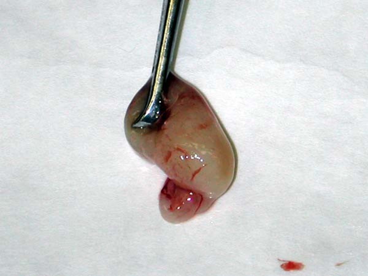
Courtesy of Dr. Michelle Woodward.
Nasopharyngeal polyps are uncommon, benign, smooth, pink, fleshy, pedunculated, inflammatory growths of connective tissue found in the external ear canals of young cats. They arise from the mucosal lining of the tympanic bulla, the pharyngeal mucosa, or the auditory tube. Although some cats may not demonstrate clinical signs associated with middle ear polyps, extension of the polyps may lead to otitis externa, otitis interna, or nasopharyngitis. These polyps may be congenital or may result from chronic bacterial otitis media, commonly found in cats with upper respiratory diseases. They are rarely found in dogs. They are not neoplastic, and viruses have not been identified in polyp tissue. Clinical signs are associated with the area of extension and are typically chronic and progressive. Clinical signs include:
head shaking
otorrhea
Horner syndrome
head tilt
ataxia
nystagmus
circling
facial nerve paralysis
nasal discharge
stertorous breathing
sneezing
Diagnosis of Nasopharyngeal Polyps
Diagnosis of nasopharyngeal polyps involves sedation and deep otoscopic examination of the horizontal canal. Purulent discharge from the bulla may need to be flushed and suctioned from the ear canal to visualize the polyp. The tympanic membrane may be intact if the polyp has not extended to the external ear. Use of a video otoscope greatly facilitates visualization and treatment of polyps. When evaluating cats for inflammatory polyps, both ear canals and the nasopharynx should be evaluated so as not to miss any potential masses. Nasopharyngeal polyps originating from the eustachian tube may be seen by retracting the soft palate rostrally. Radiography of the bullae may reveal an opacity in the affected bulla. CT or MRI may be helpful if a mass is suspected in the tympanic bulla that cannot be seen otoscopically. Definitive diagnosis is made via histopathology.
Treatment of Nasopharyngeal Polyps
There are both minimally invasive and surgical techniques to remove polyps. The most simple is traction avulsion, which may have a lower recurrence rate if performed with a lateral approach and the stalk is removed. Although this is a reasonable first-line approach, a ventral bulla osteotomy is considered the treatment of choice by some surgeons. In cats with chronic otitis and subsequent stenosis of the horizontal ear canal, this may be the only way to approach the mass. Incomplete removal of the base of the polyp by traction avulsion alone leads to rapid regrowth and return of clinical signs in 15%–50% of cats. Topical steroids in the bulla for 30–45 days seems to retard this regrowth. Systemic antibiotic therapy for the bacterial otitis media is also indicated.
For More Information
Also see pet health content regarding nasopharyngeal polyps in cats.
