

The primary functions of the urinary system include the following:
excretion of waste products of metabolism
maintenance of a constant extracellular environment through conservation and excretion of water and electrolytes
production of the hormone erythropoietin, which regulates hematopoiesis
production of the enzyme renin, which regulates blood pressure and sodium reabsorption
metabolism of vitamin D to its active form (1,25-dihydroxycholecalciferol)
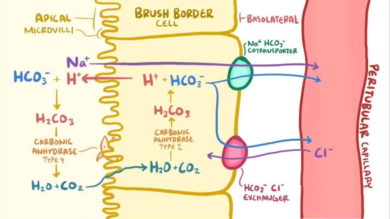
regulation of acid-base balance (see the )
excretion of drugs and toxic compounds

Diagnosis of Urinary Tract Disorders in Animals
Many abnormalities of the urinary system can be diagnosed from signalment, history, physical examination, serum biochemical profile, urinalysis, and aerobic bacterial culture of urine.
The patient's history should include information regarding changes in water consumption, frequency of urination, volume of urine produced, appearance of urine, and behavior. It is also important to obtain information about historical and current drug administration, appetite, diet, changes in body weight, and previous illnesses or injuries.
To detect disorders of the urinary system, the veterinarian should perform a complete physical examination including palpation of the bladder and examination of external genitalia. In dogs, rectal examination should be performed to evaluate the urethra in both sexes and the prostate in males. In cats, rectal examination might not be feasible because of their small size; however, the kidneys are generally easier to palpate in cats than in dogs. A full neurological examination should be performed on all patients with micturition disorders.
Additional diagnostic tests—such as CBC, blood gas analysis for acid-base status, blood pressure, urine protein:creatinine ratio, symmetric dimethylarginine (SDMA) test, survey abdominal radiography, abdominal ultrasonography, contrast studies of the upper and lower urinary tract, cystoscopic examination of the urinary bladder, and renal biopsy—can also provide valuable information.
Urinalysis
One of the most important diagnostic tests for evaluating urinary tract disorders is urinalysis. (Also see Urine Appearance.)
Urine can be collected by one of four methods: spontaneous micturition, manual compression of the urinary bladder, catheterization, and cystocentesis. Each method has advantages and disadvantages (see the table ).
Advantages and Disadvantages of Urine Collection Methods
Method of Collection | Advantages | Disadvantages |
|---|---|---|
Spontaneous micturition | No risk (eg, trauma, bacterial infection) to patient. Avoids iatrogenic hematuria. | Can contain debris (eg, bacteria, exudate) from lower urinary and genital tracts. If bacterial growth appears in urine culture, must differentiate between urethral contamination and UTI. Quantitative urine culture required. |
Manual compression of urinary bladder | Provides method to obtain urine sample when voluntary micturition has not occurred. | Potential for trauma to urinary tract, resulting in hematuria. Can be stressful for patient, especially if bladder is painful. If bacterial growth appears in urine culture, must differentiate between urethral contamination and UTI. Quantitative urine culture required. |
Catheterization | Provides method to obtain urine sample when other methods of collection have failed. | Potential for trauma to urinary tract, especially urethra. More invasive than other methods; sedation may be required. Risk of introducing bladder infection. If bacterial growth appears in urine culture, must differentiate between urethral contamination and UTI. Quantitative urine culture required. Least desirable method of urine collection. |
Cystocentesis | Preferred method of collection for urine culture. Avoids contamination of sample by debris (eg, bacteria, exudate) from lower urinary and genital tracts. Provides sterile urine sample most reliable for bacterial culture and antimicrobial sensitivity testing. | Potential risk of trauma if performed incorrectly or patient moves during procedure. Potential for iatrogenic hematuria. More invasive than spontaneous micturition. Potential for bacterial contamination of sample if needle penetrates colon during procedure. |
Abbreviations: UTI, urinary tract infection. | ||
Urinalysis results should include method of collection, urine specific gravity, color, turbidity, pH, glucose, ketones, bilirubin reagent test, occult blood, protein, and leukocytes (urine dipstick leukocyte tests are unreliable in cats). The following important factors should be taken into consideration when performing urinalysis:
Pearls & Pitfalls
|
Urine specific gravity should be obtained using a refractometer.
Microscopic examination of urine sediment should include quantitation of RBCs, WBCs, epithelial cells, renal casts, bacteria, yeast, parasitic ova, fat, sperm, and crystals.
Delay in analyzing urine samples can result in artifacts (eg, changes in urine pH, formation of crystals, etc), so it is important to note the time when the sample was collected and the time when it was analyzed.
If a sample will not be analyzed immediately, it should be refrigerated.
Protein in urine should be evaluated in light of urine specific gravity. Protein in a concentrated urine sample might not be noteworthy; however, the same amount in a dilute sample could indicate a problem.
Urine dipstick tests provide a semiquantitative assessment of protein and can be influenced by urine pH. Therefore, they should be used only as screening tests for protein, not for definitive diagnosis of proteinuria. (For a review of pH concepts, see the .)
A urine protein:creatinine ratio from a single urine sample or from a pooled 24-hour urine sample is required to quantify the amount of protein in urine. See the table .
Proteinuria Assessment Based on Urine Protein:Creatinine Concentration Ratioa
Substaging of Chronic Kidney Disease by Proteinuria Status | Urine Protein:Creatinine Concentration Ratio | |
|---|---|---|
Dogs | Cats | |
Nonproteinuric | < 0.2 | < 0.2 |
Borderline proteinuric | 0.2–0.5 | 0.2–0.4 |
Proteinuric | > 0.5 | > 0.4 |
aBased on International Renal Interest Society (IRIS) guidelines. | ||
Urine protein:creatinine ratios must be interpreted in the context of other information from the urinalysis. Inflammation and hematuria can falsely increase urine protein:creatinine ratios, although hematuria generally has minimal effects.
Bacterial Culture of Urine
Urinalysis is unreliable to exclude a urinary tract infection (UTI). Not all UTIs are associated with an inflammatory response. In addition, bacterial counts of > 10,000 rods/mL and > 100,000 cocci/mL of urine are required to consistently find bacteria in a urine sample using light microscopy. Approximately 25%–30% of all dogs with UTI have urine bacterial counts below these thresholds at the time of specimen collection, so urine culture is important for excluding UTI.
Urine samples for bacterial culture can be obtained by the same methods used to obtain samples for urinalysis; however, the preferred method is cystocentesis. Urine obtained by cystocentesis should be sterile. If urine samples are collected by methods other than cystocentesis, a quantitative urine culture should be requested. The method of urine sample collection is important for interpreting the data:
If the sample is collected by spontaneous micturition or manual compression, bacterial colony counts ≥ 100,000 CFUs/mL of urine in dogs or ≥ 10,000 CFUs/mL of urine in cats indicate the presence of clinically relevant bacteriuria consistent with UTI. Colony counts of 10,000–90,000 CFUs/mL in dogs and 1,000–10,000 CFUs/mL in cats suggest UTI.
If the sample is collected by catheterization, colony counts ≥ 10,000 CFUs/mL of urine in dogs and ≥ 1,000 CFUs/mL of urine in cats indicate the presence of clinically relevant bacteriuria consistent with UTI. Colony counts of 1,000–10,000 CFUs/mL in dogs and 100–1,000 CFUs/mL in cats suggest UTI.
Serum Biochemical Profile
Serum biochemical analysis, including measurement of concentrations of BUN, creatinine, calcium, phosphorus, bicarbonate, and serum electrolytes, is useful for diagnosing many urinary tract disorders and can provide a crude indication of glomerular filtration rate (GFR).
Although increases in BUN and creatinine concentrations suggest renal dysfunction, these values are influenced by nonrenal factors as well. For example, dehydration can lead to increases in BUN and serum creatinine concentrations that are not associated with renal failure. BUN concentration can also be influenced by diet and GI bleeding, and it is considered inferior to creatinine concentration as an indication of GFR. Serum creatinine concentrations can be falsely decreased in patients with severe muscle wasting and falsely increased in patients with severe muscle damage.
Although BUN and serum creatinine concentrations increase as GFR decreases, this relationship is not linear. Large changes in GFR early in renal disease lead to only small increases in BUN and serum creatinine concentrations, whereas small changes in GFR in advanced renal disease can be associated with large changes in BUN and serum creatinine concentrations.
SDMA concentration is not affected by the factors that affect serum creatine and BUN concentrations, and the test for SDMA can detect changes in GFR earlier than either serum creatinine or BUN concentration can. SDMA may increase with as little as 25% loss of kidney function, versus creatinine, which does not increase until 75% loss of kidney function. Therefore, SDMA is more sensitive than creatinine as an indicator of renal function.
OtherDiagnostic Tests
More sensitive methods to detect renal dysfunction include plasma clearance tests (eg, inulin clearance), radionuclide techniques, endogenous creatinine clearance, and exogenous creatinine clearance. However, these tests are impractical to perform routinely in clinical practice.
The iohexol clearance test was once an alternative to detect renal dysfunction; however, it has largely been replaced by the SDMA test. The iohexol clearance test is performed by determining the appropriate dose of iohexol based on the patient's body weight and administering this dose intravenously. Blood samples are then collected 2, 3, and 4 hours after administration to determine iohexol clearance, which is then used to measure glomerular filtration rate.
Depending on the cause of the urinary tract disorder, additional valuable information can be obtained through radiographic imaging, ultrasonographic examination, and cystoscopic examination of the bladder. Despite the presence of disease, not all portions of the kidney are always equally affected; therefore, renal biopsies can miss appreciable disease and are rarely useful when evaluating renal dysfunction. An exception is in patients with substantial proteinuria.
Blood gas analysis or serum bicarbonate concentrations provide useful information on acid-base status, especially for patients with renal dysfunction. Metabolic acidosis is a common problem with chronic kidney disease and can result in protein catabolism.
Urinary neutrophil gelatinase-associated lipocalin (NGAL) and cystatin C (CysC) have shown promise as biomarkers for early detection of renal injury in cats and dogs (1, 2, 3, 4, 5).
Principles of Treatment of Urinary Diseases in Animals
Diseases of the urinary system can result from a variety of pathological processes, and appropriate treatment depends on the location, severity, and cause of the problem. For detailed discussions, see:
Noninfectious Diseases of the Urinary System in Large Animals
Noninfectious Diseases of the Urinary System in Small Animals
Also see Systemic Pharmacotherapeutics of the Urinary System.
If a urinary tract condition is not life-threatening, appropriate diagnostic samples should be collected before treatment is initiated. Some diagnostic tests have the potential to cause substantial harm, and some treatments can make diagnosis of specific conditions more difficult. If the specific cause cannot be determined, nonspecific and supportive care (eg, monitoring the patient, administering fluids, treating acidosis, managing nutrition with an appropriate therapeutic diet) should be instituted.
For More Information
International Renal Interest Society: IRIS Guidelines
Chew DJ, DiBartola SP, Schenck PA. Canine and Feline Nephrology and Urology. 2nd ed. Elsevier/Saunders; 2011.
Bartges J, Polzin DJ. Nephrology and Urology of Small Animals. John Wiley & Sons; 2011.
References
Paes-Leme FO, Souza EM, Paes PRO, et al. Cystatin C and IRIS: advances in the evaluation of kidney function in critically ill dog. Front Vet Sci. 2021;8:721845. doi:10.3389/fvets.2021.721845
Miyagawa Y, Akabane R, Ogawa M, Nagakawa M, Miyakawa H, Takemura N. Serum cystatin C concentration can be used to evaluate glomerular filtration rate in small dogs. J Vet Med Sci. 2021;82(12):1828-1834. doi:10.1292/jvms.20-0201
Ghys LF, Paepe D, Duchateau L, et al. Biological validation of feline serum cystatin C: the effect of breed, age and sex and establishment of a reference interval. Vet J. 2015;204(2):168-173. doi:10.1016/j.tvjl.2015.02.018
Wu PH, Hsu WL, Tsai PJ, Wu VC, Tsai HJ, Lee YJ. Identification of urine neutrophil gelatinase-associated lipocalin molecular forms and their association with different urinary diseases in cats. BMC Vet Res. 2019;15(1):306. doi:10.1186/s12917-019-2048-9
Monari E, Troìa R, Magna L, et al. Urine neutrophil gelatinase-associated lipocalin to diagnose and characterize acute kidney injury in dogs. J Vet Intern Med. 2020;34(1):176-185. doi:10.1111/jvim.15645


