

A breeding soundness examination, which should take place before any dog or cat is purposefully bred, is the opportune time for veterinary input regarding good breeding practices. Because of pet overpopulation, dog and cat breeding should be left to educated and responsible breeders, to whom potential clients interested in obtaining a purebred puppy or kitten can be referred.
Breeding soundness means a dog or cat is healthy and free from heritable defects based on phenotypic evaluations such as radiography (eg, elbow dysplasia), ultrasonography (eg, renal dysplasia, tricuspid dysplasia), ophthalmoscopy (eg, cataracts), physical examination (eg, patellar luxation), or specific DNA testing (genotypic screening) when available (eg, progressive rod-cone degeneration). Several resources are available to veterinarians researching purebred dog and pedigreed cat genetic recommendations.
A thorough history should be obtained concerning past health, health of close relatives, current medications, diet, and any supplements. The appropriateness of diet, medications, and supplements for a breeding dog or cat should be discussed.
Client handouts outlining potential problems associated with breeding (dystocia, mastitis, metritis, eclampsia, prostatitis, etc) are helpful.
Potential breeding animals should be evaluated historically (eg, atopic, epileptic), physically (eg, brachycephalic syndrome, entropion, cryptorchidism), and mentally (eg, fearful, aggressive). (See Society for Theriogenology position statement on the welfare of breeding dogs.)
Female and male dogs should both undergo routine Brucella canis screening, even before the first breeding, because mucosal transmission does not require copulation; it can be transmitted through urine via oral mucosa. Subsequently, bitches should be screened for brucellosis before each cycle when a breeding is planned; stud dogs should be tested at least annually and only bred to currently negative bitches.
Kennels should prescreen any incoming dogs and quarantine appropriately (usually 2 weeks) so that breeders are kept in a Brucella-free environment. Imported or rescued dogs in particular must be screened for Brucella.
A negative B canis screening test is reliable (unless < 2–3 weeks after exposure); positive results require confirmatory testing because false-positives can occur. A negative screening test in a dog with clinical signs (eg, diskospondylitis) or history (eg, abortion, semen abnormalities, epididymitis) compatible with brucellosis should prompt additional testing. Clinicians should contact their commercial or academic laboratory for updated screening and confirmatory protocols because test availability varies. (See Society for Theriogenology position statement on brucellosis testing, revised 2024).
Laboratory screening tests currently available include culture of blood (non-EDTA; heparin or sodium citrate preferred), urine, semen, aborted fetuses/placentas and associated vulvovaginal discharge, and male and female reproductive organs. Handling of such tissues represents a biohazard; appropriate handling precautions should be observed. Positive test results are diagnostic, whereas negative results are not reliable.
Serological testing specific for B canis can have variable results, depending on circulating titers; negative results can be unreliable.
ELISA can be used for screening and confirmatory testing. (Also see Cornell University's Brucella canistest).
The agar gel immunodiffusion test is advised as a confirmatory test for suspected cases, with best results 8–12 weeks postinfection; it requires specialized media and trained personnel.
PCR assay can detect B canis in whole nonheparinized blood, urine, semen and vaginal secretions; testing is reliable but has limited availability.
Queens and toms should be screened for feline leukemia virus, feline immunodeficiency virus, and feline infectious peritonitis and housed in a virus-free cattery/home.
As part of evaluating fitness for breeding, a veterinarian should assess the general health of bitches and queens > 5 years old with a CBC, serum biochemical analysis, and urinalysis.
Visits by a veterinarian to catteries experiencing reproductive difficulties are often rewarding because basic husbandry problems can be discovered (light management, crowding, breeding management).
The Breeding Soundness Examination in Female Dogs and Cats
In bitches, in addition to a routine physical, the normalcy of the mammary glands and vestibulovaginal canal should be evaluated.
Mammae may be asymmetric in number; nipples should not be inverted (see ).
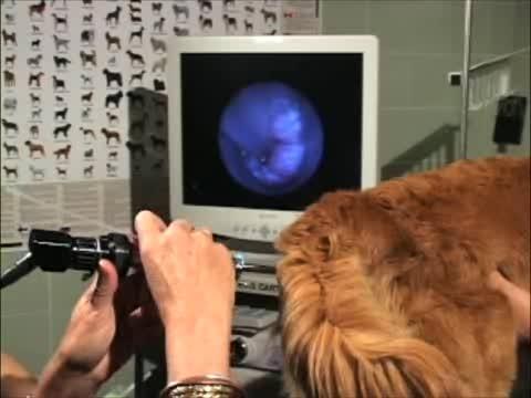
A digital vaginal examination should always be performed in the bitch before breeding; this may only be possible when a female is in estrus. Vaginoscopy provides the most thorough evaluation and can usually be performed in an estrual bitch when in proestrus (see ).
Vestibulovaginal defects (strictures) can interfere with copulation and whelping, necessitating prebreeding repair or planned artificial insemination and elective cesarean section (see ). Heritability is unknown.
See also the .


Female dog with an inverted nipple. Note accumulation of sebaceous debris and focal excoriation.
Courtesy of Dr. Autumn Davidson.


A rigid vaginoscope (cystourethroscope) positioned to illustrate the scope length necessitated by the length of the vagina in female dogs. The vaginoscope permits transcervical sampling as well as intrauterine insemination.
Courtesy of Dr. Autumn Davidson.


Orientation of the vulva, vagina, cervix, and uterus in the bitch. The position of a cotton-tipped swab for obtaining a vaginal mucosal cell sample is shown.
Courtesy of Dr. Alexander Frederick.

Vaginal strictures are congenital and occur as a septate strand or a circumferential band; they are commonly found at the vestibulovaginal junction, cranial to the urethral papilla. In some cases, the normal narrowing of the vestibulovaginal junction in the maiden (unbred) bitch could be misinterpreted as a stricture; this should be reassessed when the bitch is in estrus and vaginal tissues have become pliant (see ). Septate strands can be easily resected digitally (if thin) or surgically (if bandlike); however, circumferential strictures are difficult to resolve without episiotomy and major revision, and they tend to re-form (see ).


Endoscopic view of the vestibulovaginal junction in a dog, demonstrating normal narrowing.
Courtesy of Dr. Autumn Davidson.


Intraoperative photograph demonstrating septate vaginal strand revision performed during a vaginoscopy.
Courtesy of Dr. Autumn Davidson.
Routine prebreeding vaginal cultures are not advised because the vagina (and prepuce) normally harbors, as normal flora, a wide variety of bacteria, including beta-hemolytic streptococci and Mycoplasma spp.
Queens should also have mammary glands examined for symmetry and normalcy of nipples.
Before an anticipated breeding, female dogs and cats should be in optimal body condition to improve conception rate and whelping/queening outcome.
Proper nutrition, housing, and exercise strategies for pregnancy and lactation should be outlined. Diets designed for pregnancy and lactation are recommended, especially in queens, to provide optimal nutrition; owners should avoiding overfeeding to prevent obesity. Unnecessary supplements such as calcium can actually be problematic (predisposes to eclampsia).
Breeders commonly advise skipping cycles between breedings; this is not optimal husbandry because the inevitable exposure to estrogen (queen) and progesterone (bitch; sometimes queen) during each estrous cycle promotes cystic endometrial hyperplasia and may result in pyometra. Bitches and queens kept in optimal health can be bred sequentially and should then be considered for ovariohysterectomy or ovariectomy when no further breedings are planned, avoiding pyometra.
Bitches should be currently vaccinated for core infectious diseases (canine distemper, parvovirus, adenovirus 2, parainfluenza, influenza, and rabies). Other noncore vaccinations should be administered only according to good medical practice (appropriate for the dog’s age, health status, home and travel environment, and lifestyle).
Queens should similarly be vaccinated appropriately (based on duration of immunity recommendations) for rabies, feline panleukopenia, rhinotracheitis, and calicivirus. Vaccination against feline leukemia virus and other noncore diseases should be done when indicated by good medical practice, based on risk factors associated with the cat’s age and husbandry.
Revaccination of bitches and queens before breeding is not advised, unless their vaccination status is inadequate or unknown.
Vaccination during pregnancy is advised only when prior vaccination status is lacking or unknown and risk of exposure is high (eg, in a shelter). In that case, the use of recombinant core vaccines is optimal.
During pregnancy and lactation, the use of preventive medication for heartworm disease and internal and external parasite control (according to manufacturers’ labeling stating safety in breeding animals) is advised.
Infectious disease prevention in the pregnant female through isolation during the last third of pregnancy is important (eg, avoiding exposure to canine herpesvirus and feline upper respiratory infections).
Client education concerning normal whelping and queening events and about the timely identification of dystocia is essential. Fetal and uterine monitoring systems, developed for routine use in female dogs and cats, improve neonatal survival and decrease morbidity and mortality rates in dams.
The Breeding Soundness Examination in Male Dogs and Cats
A complete breeding soundness examination (BSE) in stud dogs and tom cats consists of a general and breeding history, physical examination, semen evaluation, and testing for B canis in the dog and feline viral leukemia, immunodeficiency and infectious peritonitis in toms.
If infertility is suggested from the history, it should be established that adequate breeding management was practiced and that bitches or queens had normal fertility when bred to other dogs or tom cats. The time sequence of when litters were sired and when bitches/queens did not conceive should be recorded, as should any recent illness during or before the time the bitches/queens were bred. These records should be assessed to determine whether infertility may be transient (eg, fever can adversely affect semen production for > 60 days after the initial insult).
A general physical exam with attention to the reproductive tract should be performed, preferably after any attempt to collect semen or on another day. altogether.
The penis and prepuce should be examined; problems such as a persistent frenulum, masses, or mucosal changes due to balanoposthitis may prevent normal intromission. Abrasions or lacerations on the penis may bleed during breeding, and blood may be present in the semen. The penis should be fully extruded from the prepuce and examined. This may require sedation in toms. If hair accumulates around the base of the feline penis (hair ring), it can prevent copulation and should be removed. A feline persistent penile frenulum, if present, will interfere with normal copulation and should be addressed with appropriate sedation and analgesia. A persistent penile frenulum may be associated with early neutering in kittens not intended for breeding and occurs less commonly in stud cats.
The scrotum, testes, and epididymides should be palpated. Scrotal skin should be visualized for dermatitis or excoriation. Small, soft testes are usually associated with poor semen quality; a greatly enlarged testis suggests orchitis or epididymitis. Masses suggesting neoplasia may also be palpable. Scrotal abnormalities such as dermatitis may adversely affect semen quality by decreasing scrotal thermoregulation. Length, width, and height of testes should be measured with blunt calipers or with ultrasonography; these measurements are often of value for future comparison in cases of suspected testicular degeneration. Additionally, total scrotal width is highly correlated with body weight and an estimate of the dog’s sperm production potential.
The canine prostate should be digitally palpated per rectum. Palpable abnormalities (pain or asymmetry) and/or semen abnormalities always warrant ultrasonographic evaluation of the prostate and further clinical testing as indicated (eg, urinalysis, urine culture).
The most common prostatic problem in mature (> 5 years old) intact dogs is benign prostatic hyperplasia (BPH); the prostate appears uniformly enlarged and is usually not painful on palpation per rectum. Dogs with BPH might be subclinically affected or have a history of hematuria and/or hemospermia or tenesmus. In dogs with clinical signs, further evaluation is warranted for prostatitis or prostatic neoplasia. Valuable breeding dogs can be treated medically for BPH with 5-alpha-reductase inhibitors, which prevent the conversion of testosterone to active dihydrotestosterone in the prostate alone.
Ultrasonography has become a common adjunctive tool for prostate evaluation in dogs, allowing for accurate measurements and assessment of the gland's echotexture for identification of potential pathological changes, in which case a fine needle or biopsy instrument can be guided to sample a lesion.
Feline prostatic disease is rare.
In cryptorchidism, a common congenital defect in males, one or both testes fails to descend into the scrotum by 6 weeks of age in the dog and by birth in the cat. Diagnosis is usually made on physical examination.
Testicles normally descend into the scrotum by 6 weeks in the dog; scrotal testes should be present at birth in the cat but can be difficult to palpate due to their small size. Descent as late as 10 months has been documented in dogs but is not considered normal.
Unilateral cryptorchidism does not result in infertility. Bilaterally cryptorchid males are sterile but have normal testosterone concentrations.
In dogs, cryptorchidism is hereditary, and affected animals should not be bred. Both late descent and failure of descent are heritable. Both parents of affected individuals should be implicated as carriers; cryptorchidism is sex-limited but not sex-linked because the gene is on the X chromosome.
Because retained testes have a higher incidence of neoplasia and subsequent torsion and the condition is heritable, bilateral orchiectomy is recommended (see ).


Postoperative image comparing intra-abdominal cryptorchid testicle having undergone malignant transformation and subsequent torsion with the normal scrotal testicle.
Courtesy of Dr. Autumn Davidson.
Attempts at inducing descent with medical therapy using gonadotropins or testosterone have been unsuccessful and are not ethical. Orchiopexy is also considered unethical.
Failure of one testis to develop (true monorchidism) is rare in dogs. Serum luteinizing hormone (LH) concentrations are high (> 1 ng/mL) if a dog or cat is completely neutered; serum antimüllerian hormone is positive in most postpubertal dogs and cats with any gonadal tissue. Examination for penile barbs in the tom cat also confirms the presence of testosterone.
A persistent penile frenulum in the dog prevents protrusion of the penis from the prepuce and thus copulation with a tie. Treatment is surgical.
Chordee (deviation of the penis) is uncommon; affected animals require assistance in breeding or may be bred via artificial insemination. Heritability is unknown.
Hypospadias (displaced urethral opening) is commonly associated with cryptorchidism. Some type of reconstructive surgery involving urethrostomy and penile amputation is usually necessary.
Phimosis can be caused by stenosis of the preputial opening, which may be congenital or result from chronic inflammation (trauma or bacterial dermatitis). Any underlying cause should be treated and then, if necessary, the opening enlarged surgically. Most of these defects have a heritable component.
Semen Evaluation
Ideally, a complete semen evaluation should be performed in male dogs intended for breeding and repeated at least annually in active stud dogs. Semen is readily collected from most dogs by manual stimulation; the presence of a teaser (estrual) female is advised to optimize results by improving libido. Semen may be collected in the absence of an estrual bitch. The pheromone methylparaben may be helpful for collection in the absence of a bitch; some veterinarians freeze swabs of estrual bitch urine or vaginal secretions for this purpose.
All equipment (artificial vagina, collecting tubes, pipettes, slides, and coverslips) should be room to body temperature, dry, and free of water and contaminants such as chemical disinfectants.
Canine ejaculate consists of 3 fractions—the first and third are of prostatic origin, while the second is sperm-rich. Sperm production is related to testicular size, so large dogs should produce higher sperm counts than small dogs.
Semen evaluation should include an assessment of libido, total sperm count per ejaculate (normal in dogs is 200–400+ million; cats 6–16 million), sperm motility (normal > 90% progressively motile, with moderate to fast speed), and morphology (> 80% normal).
Sperm count (sperm/mL) can be determined with a hemocytometer or by spectrophotometry. Sperm per ejaculate is calculated by multiplying the sperm count/mL by the total volume of sperm-rich ejaculate collected.
Motility is evaluated in an unstained sample as soon as the sample is collected, ideally using clean prewarmed slides; although computer-assisted semen analysis equipment is available, routine light microscopy (using a 40X objective) is sufficient with practice.
Several commercially available stains are suitable for morphology examination; eosin-nigrosin and Giemsa stains are used most commonly. Semen morphology slides are prepared and stained as any cytology sample; performing 100- to 200-cell counts noting morphological defects is advised.
Primary defects occur during spermatogenesis, secondary defects occur during sperm transport and storage, and acquired defects occur secondary to inflammatory genitourinary disease, thermoregulatory disorders of the testes, heat stress, drugs, hematocele, hydrocele, and immune-mediated orchitis. Defects are also categorized as major or minor. Iatrogenic defects can be caused by stains in slide preparation (see ).


Photomicrograph of a sperm morphology slide demonstrating teratospermia; a microcephalic spermatozoon is shown next to 3 normal spermatozoa. This is a major morphological defect affecting sperm function. Commercial dark blue staining and fixative solution in buffered salt; original magnification, 1000X.
Courtesy of Dr. Autumn Davidson.
An adequate amount of the third fraction should be collected to ensure that the entire sperm-rich fraction has been acquired and to permit evaluation of the prostatic component, which should be clear (free of urine and cellular contamination). Subfertility or infertility should never be diagnosed based on one collection. Repeat collections > 48 hours apart should be performed. If the sample is aspermic, semen ALP can be measured in the ejaculate to assess whether the ejaculate was complete, because semen ALP is an epididymal marker.
Concentrations > 5,000 mcg/dL indicate the ejaculate included the second, normally sperm-rich fraction. Concentrations < 5,000 mcg/dL indicate either bilateral obstructive disease or libido problems preventing the release of the second fraction. Sperm function (eg, capacitation and acrosome reaction, zona pellucida binding) is not assessed with routine semen evaluation.
Collection of semen for evaluation is difficult in toms, unless the cat has been trained to ejaculate into an artificial vagina, chemical ejaculation is undertaken, or electroejaculation equipment is available. Cats can be trained to ejaculate with manual stimulation in some instances, but training can take weeks to months. Chemical ejaculation, using urethral catheterization under dexmedetomidine sedation or fine-needle aspiration of the testes to assess sperm presence and cytology, has been described. Nonspecific methods of evaluating a tom for spermatogenesis include assessing the urine for sperm or collecting a vaginal wash from the queen immediately after copulation; spermatozoa disappear from the vagina within 1–2 hours of copulation.
Semen is best collected in a disposable artificial vagina, which can be closed or open-ended and connected to a plastic tube (see ). A sterile nonspermicidal lubricant can be used but is usually not necessary and best used after the collection. The prepuce is gently pushed caudal to the bulbus glandis while the artificial vagina is slipped over the cranial penis. As soon as the bulbus glandis is exteriorized from the prepuce and is within the artificial vagina, the shaft of the penis is gently massaged through the cone, with gentle pressure applied immediately caudal to the bulbus. Care must be taken to avoid full erection of the bulbus glandis within the prepuce, as pain will result. Constant pressure is maintained; erection, and eventually ejaculation, should be achieved. If a teaser bitch is present, mounting is permitted; the artificial vagina prevents actual copulation.


A) The male dog is mounting a toy dog swabbed with estrual scent; note nonskid rug in use. B) The veterinarian has inserted the artificial vagina over the erect, extruded penis while the dog is thrusting. C) The second fraction (white) of semen is collected into a plastic tube. In this case, a latex artificial vagina is in use.
Courtesy of Dr. Autumn Davidson.
For sample recovery and analysis, warm saline solution (0.1–0.2 mL) is flushed into the queen's vagina and aspirated, the sample is centrifuged, and the sediment examined (new methylene blue or routine hematologic stains are adequate). This analysis should not be performed if the breeding is desirable, but only as a test of ejaculation. Breeding a questionable tom to a proven queen may be the most practical way to assess his fertility. Adequate coital contact to induce the LH surge and ovulation should be confirmed by measuring progesterone concentrations in the queen 1–2 weeks after breeding.
The first (prostatic, clear) fraction and the second (sperm-rich, cloudy) fraction should be collected (see ). After these fractions are ejaculated, close inspection of the collection tube should demonstrate that clear (prostatic) fluid is starting to layer on the cloudy second fraction; at this point, the collection may be stopped. The dog may continue to ejaculate prostatic fluid for up to 10 minutes before the erection subsides. This is when lubrication should be applied to the exposed penile mucosa. The prepuce should be examined after the penis is retracted to ensure that the entire penis is situated normally within the prepuce and that no hair is caught within the preputial orifice.
Paraphimosis may occur if the prepuce rolls inward as the penis retracts (see ).


First fraction (clear) and second fraction (white) of canine semen.
Courtesy of Dr. Autumn Davidson.


A) Erect penis after semen collection; after detumescence, the penis should slide completely within the prepuce. B) Application of nonspermicidal lubricant to facilitate detumescence and prevent paraphimosis.
Courtesy of Dr. Autumn Davidson.
Semen evaluation consists of determination of appearance, volume, concentration, motility, and percentage of morphologically normal sperm. Yellow, brown, or red samples may indicate the presence of blood or urine in the ejaculate. The volume is variable, ranging from < 2 to > 20 mL, depending on how much prostatic fluid was collected and the size of the male. The second, sperm-rich fraction is usually 0.5–2 mL.
Sperm motility should be evaluated immediately using warmed equipment (commercially available slide warmer); normally, > 70% are progressively motile (see ). Motility can be improved if the sample is centrifuged to remove any prostatic fluid and reextended in appropriate semen-extending solution.
At least 80% of the sperm should be morphologically normal. This can be observed casually in the same slide, by preparing a slide with appropriate morphology stain, or by using commercially available semen evaluation machinery.
The sperm concentration can be determined using a hemocytometer. The total number of sperm in the ejaculate is calculated as volume × concentration. Total sperm number in the ejaculate ranges from 400 × 106 to > 1,000 × 106 and is correlated with body size. As a general rule, a dog should produce approximately 22 × 106 sperm/kg (10 × 106 sperm/lb).

Light microscope, slide warmer (Vetko-2) , centrifuge (LW Scientific), and automated semen evaluator (Sperm cBlue Mini tube).
Courtesy of Dr. Autumn P. Davidson.
Any dog intended for breeding, and every dog investigated for infertility, should be screened for B canis.
Sperm quality may be normal or abnormal, or sperm may be absent in the ejaculate. Infertility is rare in dogs with a normal sperm evaluation, and in these cases, the history should be reviewed for mismanagement or bitch infertility. The presence of WBCs or RBCs in the ejaculate suggests inflammation of the reproductive tract, most commonly prostatitis; culture of prostatic fluid and appropriate treatment may help improve fertility.
If sperm quality is abnormal, the history should again be reviewed to determine whether the dog has been sick or stressed recently, in a hot environment, or on medications that can impair fertility (eg, ketoconazole).
Azoospermia is relatively common in dogs. It may be due to failure of the dog’s testes to produce sperm or to failure of the sperm to exit the testes because of epididymal blockage or incomplete ejaculation. The ejaculate can be tested for the presence of ALP, which is secreted by the epididymis. A high ALP value (5,000–40,000 IU/L) indicates fluid from the epididymis was collected and thus is consistent with true azoospermia. Low values (< 5,000 IU/L) suggest epididymal blockage or incomplete ejaculation; semen collection should be repeated, using a strong stimulus, such as a bitch in estrus.
Cystocentesis can be performed after semen collection to determine whether retrograde ejaculation is occurring. Swab samples of the vagina of a bitch after natural breeding may also be collected to determine whether sperm have been ejaculated; some dogs may not ejaculate with manual collection but do have normal ejaculation during natural breeding.
A dog is considered satisfactory for breeding if all the above findings are within normal limits and the dog is negative for B canis. Breeding dogs should be retested annually for B canis.
For More Information
Position statement: Brucella canis testing. Society for Theriogenology.
Brucella multiplex testing for dogs. Cornell University College of Veterinary Medicine Animal Health Diagnostic Center.
Embark Veterinary. DNA testing.
Canine health information center. Orthopedic Foundation for Animals.
DNA tests by species. UC Davis Veterinary Genetics Laboratory.
Wisdom Panel. Pet DNA kits.
Inherited disorders in cats. International Cat Care.


