


Hendra virus infection is a viral disease in Australia that can cause acute, fatal respiratory and/or neurological signs in horses of all ages. Humans and dogs have been infected as spillover events from close contact with infected horses. Definitive diagnosis is by virus isolation, PCR assay, or serological testing. There is no effective treatment for infected animals, although a vaccine can prevent clinical signs in horses.
Hendra virus was first described in 1994, after an outbreak of acute respiratory disease at a Thoroughbred training stable in Australia. In this incident, horses and one human were fatally infected. Sporadic cases continue to occur in eastern Australia, typically presenting as acute febrile illness and rapidly progressing with variable system involvement, notably acute respiratory and/or severe neurological disease. To date, more than 90 horses are known to have been infected, all of which have either died as a direct result of their infection or been euthanized on animal welfare grounds. In two separate instances of fatal Hendra virus infection in horses, a dog on the same horse property became infected but showed no clinical signs. Fruit bats of the genus Pteropus (family Pteropodidae), colloquially known as flying foxes, were determined to be the reservoir of the virus and the putative source of infection for the horses. The virus is likely transmitted from flying foxes to horses, from horses to horses, and, rarely, from horses to humans.
Hendra virus is a biosafety level 4 agent (defined as posing a high risk of life-threatening disease in humans, in accordance with the WHO Laboratory Biosafety Manual), and the use of safe work practices and personal protective equipment is essential for managing the zoonotic risk.
The earlier names of "equine morbillivirus" and "acute equine respiratory syndrome" are no longer appropriate for this disease.
Etiology and Pathogenesis of Hendra Virus Infection in Horses
Hendra virus is a large, pleomorphic, enveloped RNA virus. Although initially considered more closely related to members of the genus Morbillivirus than to other genera in the family Paramyxoviridae, subsequent studies showed limited sequence homology and negligible immunological cross-reactivity with other Orthoparamyxovirinae subfamily members. Hendra virus is genetically and antigenically closely related to Nipah virus, with which it shares > 90% amino acid homology. Both viruses have been classified in a new genus, Henipavirus, in the family Paramyxoviridae. In 2012, Cedar virus was identified in Australia and added to the Henipavirus genus. Its genome is very similar to those of Hendra virus and Nipah virus; however, Cedar virus did not cause clinical signs in experimentally challenged animals.
Evidence indicates that Hendra virus strain variation is minimal and that clinical presentation and pathology more likely vary with route of infection. In 2020, a previously unrecognized variant of Hendra virus (HeV-var) was identified as a second genotype lineage (HeV-g2); infection with this variant was clinically indistinguishable from prototypical Hendra virus infection (1). Since then, an updated quantitative PCR assay has been developed for routine surveillance, resulting in subsequent case detection.
Historically, interstitial pneumonia of variable severity was the principal finding in naturally infected horses. Similar findings were also observed in experimentally infected horses exposed by respiratory or parenteral routes.
Hendra virus has a specific tropism for vascular tissues, regardless of route of challenge. In early infection, vascular lesions may include edema and hemorrhage of vessel walls, fibrinoid degeneration with pyknotic nuclei in endothelial and tunica media cells, and numerous giant cells (syncytia) in the endothelium and sometimes the tunica media of affected vessels (both venules and arterioles). The virus becomes more widely distributed in various tissues throughout the body as infection progresses, presumably as a result of a leukocyte-associated viremia. Virus has been demonstrated in the vascular endothelium of subarachnoid and cerebral vessels and in the vasculature of the renal glomerulus and pelvis, lamina propria of the stomach, spleen, various lymph nodes, and myocardium.
With respiratory disease, progressive destruction of alveolar walls occurs, and alveolar and intravascular macrophages appear. In addition to its vascular tropism, Hendra virus can also be neurotropic, causing neuronal necrosis and focal gliosis. A feature of a 2008 outbreak at an equine veterinary clinic in Australia was severe neurological disease and an absence of respiratory disease. Various neurological signs have been observed more often in cases since approximately 2006. Thus, Hendra virus should no longer be regarded as causing predominantly respiratory disease in horses.
Epidemiology and Transmission of Hendra Virus Infection in Horses
Naturally occurring disease caused by Hendra virus has been reported only in horses, dogs, and humans. Experimentally, disease has been produced in cats, hamsters, ferrets, monkeys, pigs, and guinea pigs, but not in mice, rats, rabbits, or chickens. The clinical response and pathological findings in cats are very similar to those observed in horses.
Hendra virus infection and disease in horses has only been reported in Australia, and events are sporadic and infrequent; only 67 events were recorded from 1994 to 2023, most of which were infections of a single horse. However, during 2011–2017, the frequency of infection in horses increased (18 incidents in 2011, 8 yearly in 2012–2013, 4 yearly in 2014–2015, 1 in 2016, and 4 in 2017); geographical locations ranged from north Queensland to north New South Wales (2).
Further research summarized that these increased incidents in horses could be due to greater public awareness in reporting the disease in consideration of the risk to human health; however, environmental and ecological factors that altered the behavior of flying fox populations could also have contributed to this increased number of cases and extended geographical occurrence.
Experimentally, attempted transmission of Hendra virus from infected horses to in-contact horses or cats has been unsuccessful. Nonetheless, the possibility of respiratory transmission cannot be excluded. The frothy nasal discharge (originating from the lungs) sometimes observed terminally in naturally infected horses could plausibly provide a source of virus for aerosol transmission. Hendra virus has been found not only in nasal secretions of naturally infected horses and dogs but also in urine, blood, and oral secretions (3, 4). Available field and laboratory data seem to indicate that infection of humans or animals requires direct contact with virus-infective secretions (lung exudates), excretions (urine), body fluids, or tissues. Although Hendra virus appears to have limited infectivity, the case fatality rate among infected individuals is high: 75% in horses, 57% in humans (5).
The incubation period for Hendra virus infection in horses is 4–20 days.In 80% of known equine cases, the incubation period was ≤ 12 days; in 95%, disease developed in ≤ 15 days (5, 6, 7).
Epidemiological, serological, and virological evidence implicates fruit bats as the natural reservoir of Hendra virus (8). Information on the geographic distribution of fruit bats (flying foxes) in Australia is available from the Australian Department of Climate Change, Energy, the Environment and Water. Serological surveys have revealed a high prevalence of neutralizing antibodies in wild-caught fruit bats (Pteropus spp) in Australia and Papua New Guinea (9). The geographical distribution of the virus in fruit bats appears to be limited to Australia and Papua New Guinea, although a transition of Hendra-like to Nipah-like viruses may occur beyond Australia. Infection in fruit bats (either natural or experimental) causes no evident disease. Field and experimental evidence supports vertical transmission; isolates have been recovered from the uterine fluid and fetal tissues of a grey-headed flying fox (P poliocephalus) and a black flying fox (P alecto).
The infrequent occurrence and sporadic nature of equine cases suggest that exposure of horses to Hendra virus is, at least in part, a chance event. The modes of transmission between bats, and from bats to horses, are uncertain, as are factors that may facilitate spillover. Hendra virus has been identified in birthing fluids, placental material, aborted pups, and urine of naturally infected fruit bats and in urine of experimentally infected fruit bats. Although the exact route of transmission is unknown, horses may become infected through contact with food or water contaminated with body fluids or excretions from infected fruit bats, or through droplet inhalation via the nasal route. Risk factors for horses, such as grazing behavior (see horse grazing image), individual horse personality, large respiratory tidal volume, and highly vascularized upper respiratory epithelium, can contribute to their vulnerability to Hendra virus infection.

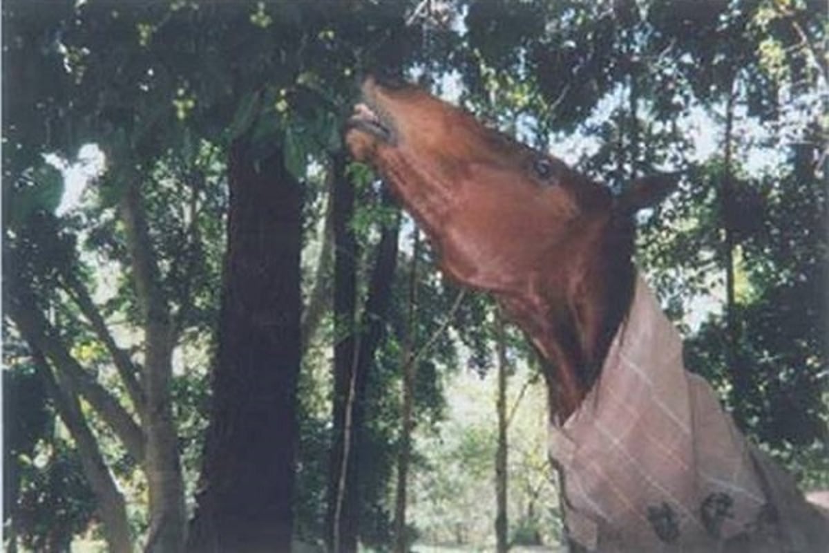
Courtesy of Biosecurity Queensland.
Clinical Findings of Hendra Virus Infection in Horses
Because of its affinity for endothelial cells, Hendra virus can cause a range of clinical signs in horses. The predominant clinical presentation may depend on which organ system sustains the most severe or compromising endothelial damage.
Hendra virus infection should be considered when there is acute-onset fever and rapid progression to death, possibly associated with severe respiratory or neurological signs; however, the absence of these signs should not preclude consideration of Hendra virus. Infection is not always fatal; in 25% of known cases, the horses recovered with detectable antibodies.
Associated respiratory signs can include the following:
pulmonary edema and congestion
respiratory distress (tachypnea)
terminal nasal discharge, which may be clear initially and progress to stable white or blood-stained froth
Associated neurological signs can include the following:
ataxia
altered consciousness (apparent loss of vision in one or both eyes, aimless walking in a dazed state)
torticollis
circling
muscle twitching (myoclonic spasms, which have been observed in acutely ill and recovered horses)
urinary incontinence
recumbency with inability to rise
terminal weakness
collapse
Other possible clinical signs include depression, severe tachycardia, facial edema, muscle trembling, anorexia, congestion of oral mucous membranes, colic-like signs (generally quiet abdominal sounds on auscultation of the abdomen in preterminal cases), and stranguria in both males and females. Proximity to fruit bat roosts or feeding sites should increase suspicion.
Where horses are paddocked, Hendra virus infection usually involves a single sick or dead horse and no transmission to in-contact companion horses. However, on several occasions, one or more companion horses have become infected after close contact with the index case before or at the time of death.
Where horses are stabled, virus transmission appears to be via direct contact with infectious body fluids or indirectly through contact with contaminated fomites, sometimes via inadvertent human-assisted transfer. To date, Hendra virus infections in stables have resulted in multiple horses becoming infected, apparently as a result of an infected horse in a paddock or outside yard being brought into the stable.
Lesions in Hendra Virus Infection
The presence of large endothelial syncytial cells on histological examination of tissue is characteristic of Hendra virus infection. Although most prominent in pulmonary capillaries and arterioles, these cells are also present in other organs (lymph nodes, spleen, heart, stomach, kidneys, and brain). Widespread fibrinoid degeneration of small blood vessels occurs in many organs, including the lungs, heart, kidneys, spleen, lymph nodes, meninges, GI tract, skeletal muscle, and bladder.
Immunohistochemical staining can demonstrate antigen specific for Hendra virus in vascular lesions and along alveolar walls. Intracytoplasmic viral inclusion bodies can be observed in infected endothelial cells by electron (but not light) microscopy.
When respiratory disease is predominant, the principal gross lesions are severe pulmonary edema and congestion and marked dilatation of subpleural lymphatics. Airways are filled with thick froth, which is often blood-tinged. Additional lesions occurring in some affected horses include increased pleural and pericardial fluids, congestion of lymph nodes, hemorrhages in various organs, and slight jaundice. Microscopically, primary lesions are those of acute interstitial pneumonia. Severe vascular damage, manifesting as serofibrinous alveolar edema, hemorrhage, capillary thrombosis, alveolar wall necrosis, and alveolar macrophages, is evident in the lungs.
When neurological disease is predominant, lesions of nonsuppurative meningitis or meningoencephalitis, including perivascular cuffing, neuronal degeneration, and focal gliosis, may be observed.
Diagnosis of Hendra Virus Infection in Horses
Clinical evaluation
PCR assay
Serological testing
Immunohistochemistry
Virus isolation
Hendra virus infection should be considered when there is acute-onset fever and rapid progression to death; however, a nonfatal outcome should not preclude consideration of Hendra virus infection.
Definitive diagnosis is based on laboratory examination of appropriate specimens to detect virus, viral antigen, viral nucleic acid, or specific antibodies. The number and type of specimens collected should be determined after a careful risk analysis by the veterinarian, to prevent human exposure to the virus. The approach to specimen collection should reflect the serious zoonotic potential of Hendra virus and should incorporate appropriate measures to avoid human exposure. Factors such as personal protective equipment availability, training, and prior experience should be considered. Minimum recommended samples include blood samples (EDTA and serum) and nasal, oral, and rectal swabs. Samples can be taken from both live and dead horses. Necropsy specimens, both fresh and fixed in 10% formalin, of lung, kidney, spleen, liver, lymph node, and brain tissue increase the likelihood of a conclusive diagnosis but also potentially increase the risk of human exposure.
Pearls & Pitfalls
|
PCR assay detects fragments of the Hendra virus genome. A positive result indicates only the presence of viral genome in the sample; it does not indicate that the virus is viable and infectious.
Serological tests, including ELISA and virus neutralization, detect antibodies against Hendra virus. ELISA is used for screening, whereas virus neutralization testing of acute and convalescent sera collected 2–4 weeks apart is confirmatory. In an unvaccinated horse, a positive titer in acute serum samples is diagnostic for recent infection. An increasing titer on the convalescent sample, whether the horse was previously vaccinated or not, also indicates recent infection.
The presence of characteristic vascular lesions on histological examination is highly suggestive of infection; lesion specificity can be confirmed by immunochemical labeling with Hendra virus reference antiserum.
Hendra virus can be isolated in a range of cell lines; Vero cells are the cell line of choice. Viral cytopathic effect, which typically develops after 3 days, is characterized by syncytia formation in infected cells. Virus isolation and other diagnostic tests involving live virus should only be attempted under biosecurity level 4 laboratory conditions. Virus isolation is not routinely performed for diagnosis, unless further epidemiological or viral strain characterization is necessary.
African horse sickness can clinically mimic Hendra virus infection and should be considered in the differential diagnosis. Other causes of sudden death that must be excluded include anthrax, botulism, equine influenza, peracute equine herpesvirus 1 infection, certain bacterial infections (eg, pasteurellosis), snakebite, and plant or chemical poisoning.
Treatment, Prevention, and Control of Hendra Virus Infection in Horses
Vaccination
Supportive care (for infected horses)
There is no specific antiviral treatment for Hendra virus infection. Commonly, infected horses with acute onset of severe clinical signs are euthanized to prevent suffering, on animal welfare grounds. For infected horses with mild clinical signs, treatment is mainly supportive to help relieve signs and reduce complications.
A vaccine, containing a noninfectious protein component (G protein) of the virus with adjuvant to stimulate immune response, has been developed; it does not contain live or inactivated virus. Introduced in 2012 and fully registered in 2015, the vaccine is available through accredited veterinarians in Australia. Healthy horses can be vaccinated from age 4 months onward with two primary doses administered 3–6 weeks apart, followed by a third dose given 6 months after the second primary dose. Boosters are administered every 12 months. Initial vaccine is delayed until age 6 months in foals born to vaccinated mares. Pregnant mares should not be vaccinated during the first 6 weeks of pregnancy or during the 2 weeks prior to the expected foaling date.
Other means of prevention focus on minimizing contact of horses with fruit-bat body fluids or contaminants and include simple, practical measures such as the following:
placing feed and water containers under cover
keeping horse paddocks mostly or entirely free of trees and shrubs (fruiting and/or flowering) that serve as food sources for fruit bats
excluding horses from the vicinity of such trees and shrubs
Control measures include the following:
euthanasia and deep burial of infected horses
monitoring, isolating, and restricting movement of in-contact animals
disinfection of potentially contaminated surfaces
A risk assessment must be undertaken to determine appropriate infection controls before personnel make close contact with an infected animal. The risk assessment should take into account animal welfare, risks to human health, and wishes of the owner. If transmission risk and other issues can be safely managed, veterinary management of the animal may continue under the control of the jurisdictional chief veterinary officer; movement control should be in accordance with the Australian Veterinary Emergency Plan (AUSVETPLAN) response strategy for Hendra virus (version 5.0, 2021).
If serological test results for a close-contact animal are positive but PCR assay results are negative, the animal is considered low risk and quarantine is not necessary. Serologically positive animals without clinical disease could also be identified during routine serological testing (eg, during export testing), and if the animal has not been vaccinated previously against Hendra virus, this result may reflect recovery from natural Hendra virus infection. Such animals are also considered low risk and are managed similarly to vaccinated animals; quarantine and movement restriction are not required.
After infected dogs have developed neutralizing antibodies, they no longer pose a transmission risk. During the acute disease phase, infected dogs need to be isolated until PCR assay results are negative and antibody tests results are positive. Although cats have only shown clinical signs experimentally, naturally infected cats would also constitute a transmission risk and would require the same stringent biosecurity measures as infected dogs.
Zoonotic Risk of Hendra Virus Infection in Horses
Hendra virus infection in humans has a 57% case fatality rate. All infections in humans have resulted from handling infected horses (both live horses and horses at necropsy), so great care must be taken to ensure the personal safety of all humans in contact with suspected or confirmed equine cases. Neither bat-to-human nor human-to-human transmission has been recorded.
Protocols to minimize risk for human exposure should be implemented on suspicion of Hendra virus infection in a horse; implementation should not wait for confirmation of infection. The approach developed by the Queensland government's Department of Primary Industries and available on the Business Queensland website is summarized here.
Veterinary practices should devise a plan in advance for how to manage Hendra virus risks. Essential directives for management include the following:
Take precautions upon suspicion of Hendra virus infection; do not wait for confirmation of infection.
Isolate sick or dead horses from humans and all other animals, including pets.
Limit human contact with in-contact horses to only essential staff.
Promote personal hygiene, especially handwashing and showering, among in-contact staff.
Identify hazards and take steps to minimize risks associated with such hazards (eg, to avoid generating splashes and aerosols, do not use a high-pressure hose when decontaminating an area).
Inform humans at risk for exposure (eg, horse owners, handlers, other veterinarians, veterinary assistants) of these risks and the procedures to be followed to minimize them.
Refer to relevant animal health and public health authorities.
In addition, the following precautions should be taken:
Personal protective equipment must be worn to protect all exposed skin, mucous membranes, and eyes from direct contact and to prevent the inhalation of airborne particulates.
Regular handwashing and washing of exposed skin with soap should be promoted.
In particular, tissues and body fluids (especially blood, respiratory and nasal secretions, saliva, and urine) from sick or dead horses should be considered potentially infectious, and appropriate precautions should be taken to prevent direct contact with, splashes from, or accidental inoculation with them.
Key Points
Hendra virus infection is a zoonotic disease affecting horses, humans, and dogs.
Fruit bats (flying foxes) in Australia are the natural reservoirs for Hendra virus.
Hendra virus transmission is from fruit bats to horses, and from horses to humans and dogs.
There is no transmission from fruit bats to humans or from human to human.
Personal hygiene and protective equipment are essential for all humans treating sick horses to protect them from infection.
For More Information
Animal Health Australia. Response Strategy: Hendra Virus (Version 5.0). Australian Veterinary Emergency Plan (AUSVETPLAN), 5th ed. 2021.
Business Queensland, Queensland Government. Hendra virus.
NSW Department of Primary Industries and Regional Development, NSW Government.Hendra virus.
Australian Veterinary Association. Guidelines for Veterinary Personal Biosecurity. 3rd ed. January 2017.
Timmiss LA, Martin JM, Murray NJ, et al. Threatened but not conserved: flying-fox roosting and foraging habitat in Australia. Aust J Zool. 2021;68(6):226-233.
Also see pet owner content regarding Hendra virus infection in horses.
References
Annand EJ, Horsburgh BA, Xu K, et al. Novel Hendra virus variant detected by sentinel surveillance of horses in Australia. Emerg Infect Dis. 2022;28(3):693-704. doi:10.3201/eid2803.211245
Summary of Hendra virus incidents in horses. Business Queensland. Last reviewed December 14, 2023. https://www.business.qld.gov.au/industries/service-industries-professionals/service-industries/veterinary-surgeons/guidelines-hendra/incident-summary
Kirkland PD, Gabor M, Poe I, et al. Hendra virus infection in dog, Australia, 2013. Emerg Infect Dis. 2015;21(12):2182-2185. doi:10.3201/eid2112.151324
Middleton DJ, Riddell S, Klein R, et al. Experimental Hendra virus infection of dogs: virus replication, shedding and potential for transmission. Aust Vet J. 2017;95(1-2):10-18. doi:10.1111/avj.12552
Halpin K, Rota PA. A review of Hendra virus and Nipah virus infections in man and other animals. In: Sing A, ed. Zoonoses: Infections Affecting Humans and Animals. 2nd ed. Springer; 2023:1493-1508. doi:10.1007/978-3-031-27164-9_40
Khusro A, Aarti C, Pliego AB, Cipriano-Salazar M. Hendra virus infection in horses: a review on emerging mystery paramyxovirus. J Equine Vet Sci. 2020;91: 103149. doi:10.1016/j.jevs.2020.103149
Animal Health Australia. Response Strategy: Hendra Virus (Version 5.0). In: Australian Veterinary Emergency Plan (AUSVETPLAN). 5th ed. 2021.
Field HE. Hendra virus ecology and transmission. Curr Opin Virol. 2016;16:120-125. doi:10.1016/j.coviro.2016.02.004
Boardman WSJ, Baker ML, Boyd V, et al. Seroprevalence of three paramyxoviruses; Hendra virus, Tioman virus, Cedar virus and a rhabdovirus, Australian bat lyssavirus, in a range expanding fruit bat, the Grey-headed flying fox (Pteropus poliocephalus)." PloS One. 2020;15(5):e0232339. doi:10.1371/journal.pone.0232339


